
The hip joint is fundamental to human movement, enabling us to walk, run, and carry out countless daily activities. As the most proximal of the lower extremity joints, the hip plays a vital role in weight-bearing and provides a wide range of motion. This detailed Muscle and Motion article will explore the hip joint’s anatomy and functions and how it plays an integral role in human movement.
Anatomy of the hip joint
The hip is a ball-and-socket joint where the rounded head of the femur fits snugly into the acetabulum of the pelvis. This structure allows multiple movements, including flexion, extension, abduction, adduction, and internal and external rotation.
Ligaments and capsule structure
The hip joint is surrounded by a strong fibrous capsule that forms a sleeve around the acetabulum and the neck of the femur. The key ligaments that provide stability are:
- Iliofemoral Ligament: Shaped like a Y, this ligament is crucial for maintaining an upright posture by limiting hyperextension. It is particularly important for standing and supporting individuals with spinal cord injuries.
- Pubofemoral Ligament: Stretching across the joint medially and inferiorly, this ligament limits hyperextension and abduction.
- Ischiofemoral Ligament: Located on the posterior aspect of the joint, this ligament prevents excessive internal rotation and adduction when the hip is flexed.
These ligaments are strategically attached in a spiral, allowing them to be flexible during leg flexion and tight during extension. This mechanism enables standing without muscle effort, instead relying on the iliofemoral ligament, which is especially beneficial for individuals with paralysis from spinal cord injuries.
Acetabular labrum
The acetabular labrum is a fibrocartilaginous structure that surrounds the rim of the acetabulum, forming a ring around the socket of the hip joint. This ring significantly deepens the socket, increasing its concavity and creating a more secure and stable fit for the femoral head. Functionally, the labrum is vital in maintaining the joint’s overall stability by preventing dislocation and enhancing the distribution of joint forces. It also plays a crucial role in sustaining intra-articular pressure, helping to seal the hip joint and retain the lubricating synovial fluid. The labrum’s unique structure also absorbs shock, protecting the joint from damage.
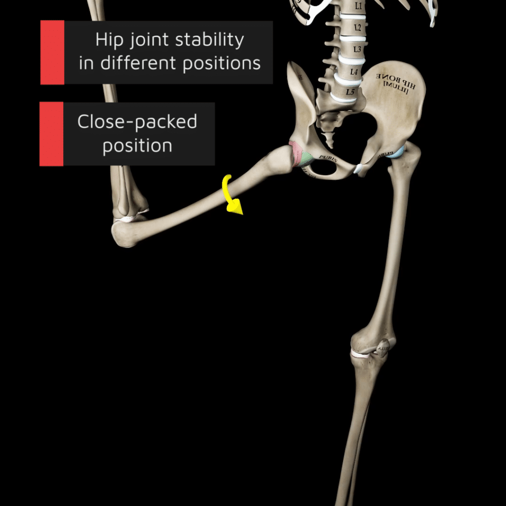
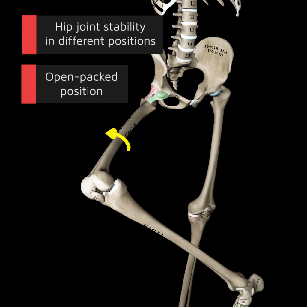
Hip joint stability and positioning
- Close-Packed Position: The hip joint achieves its highest stability when close-packed, characterized by the hip being fully extended, abduction, and internally rotated. In this state, the hip ligaments are taut, and the joint’s bony anatomy aligns to provide maximal stability, securely holding the femoral head within the acetabulum. This positioning creates tension in the ligaments and is crucial for weight-bearing activities like standing upright. This position also limits excessive movement, protecting the joint from dislocations and injuries.
- Open-Packed Position: The open-packed position occurs during hip flexion, abduction, and external rotation. This position, known as the “hook-lying” position, relaxes the hip ligaments, reducing the compressive forces on the joint and increasing the joint capsule’s laxity. However, while offering improved flexibility, the open-packed position sacrifices some joint stability due to the loose ligamentous and capsular structures.
Femoral angles and their impact
Two critical angles in the femur play a substantial role in determining hip joint function and lower limb alignment: the angle of inclination and the angle of torsion.
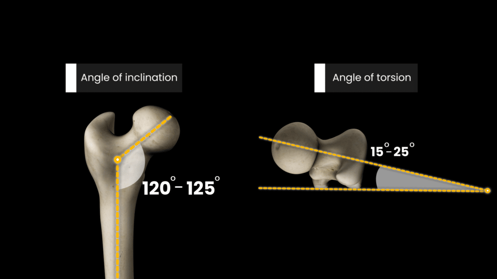
Angle of inclination
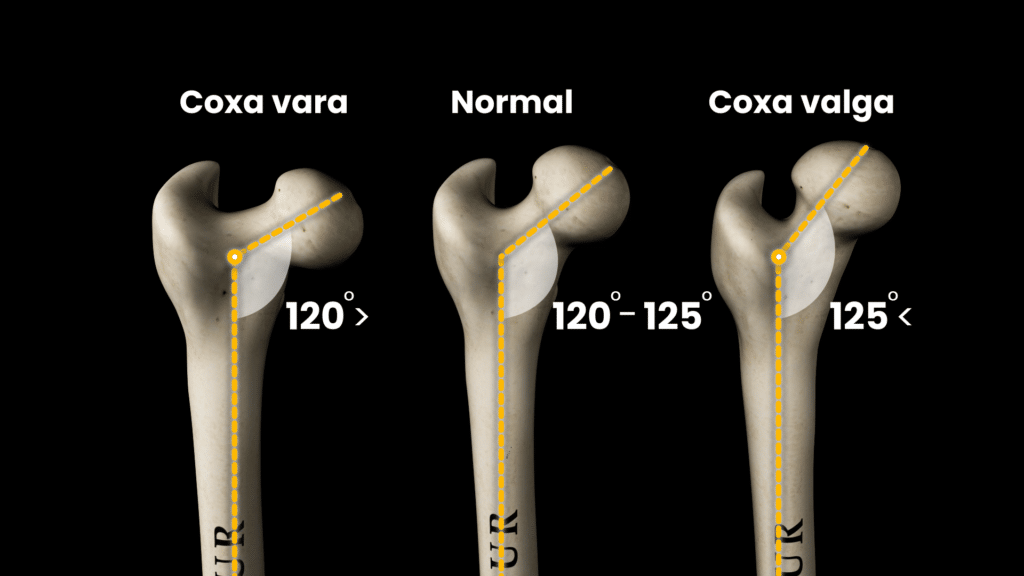
The angle of inclination, created between the femoral neck and the femoral shaft, generally ranges from 120 to 125 degrees in adults. This angle is essential because it influences how the femoral head sits within the acetabulum and how weight is distributed across the hip joint.
- Coxa Valga: An angle above 125 degrees, leading to a straighter femoral neck.
- Coxa Vara: An angle below 120 degrees, resulting in a more horizontal femoral neck
Angle of torsion
The femoral angle of torsion is the outward twist of the femoral head and neck from the shaft and typically ranges between 15 to 25 degrees. When viewed from above, the femoral head and neck align over the shaft, marked by a line through the distal femoral condyles. This angle is crucial as it dictates the hip’s rotation and, consequently, the walking pattern.
- Anteversion: An increase in this angle results in inward-facing hips, often causing a “toed-in” gait.
- Retroversion: Decreased angle leads to outward-facing hips and a “toed-out” gait.
Hip joint range of motion
Understanding the normal ranges of motion for the hip joint is crucial for assessing joint health and function. Here’s a detailed look at the typical active range of motion for the hip:
- Flexion – 120 degrees
- Extension – 10 degrees
- Abduction – 45 degrees
- Adduction – 25 degrees
- Internal rotation – 15 degrees
- External rotation – 35 degrees
These values represent the active range of motion, meaning they describe how far the joint can move by muscle action alone without any external assistance.
Muscle function and movement
The hip muscles play a pivotal role in generating and controlling the movements of the joint:
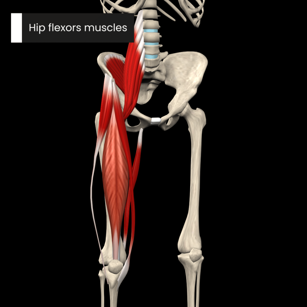
Hip flexor muscles
- Psoas major
- Psoas minor
- Iliacus
- Rectus femoris
- Sartorius
- Tensor fasciae latae
- Gracilis
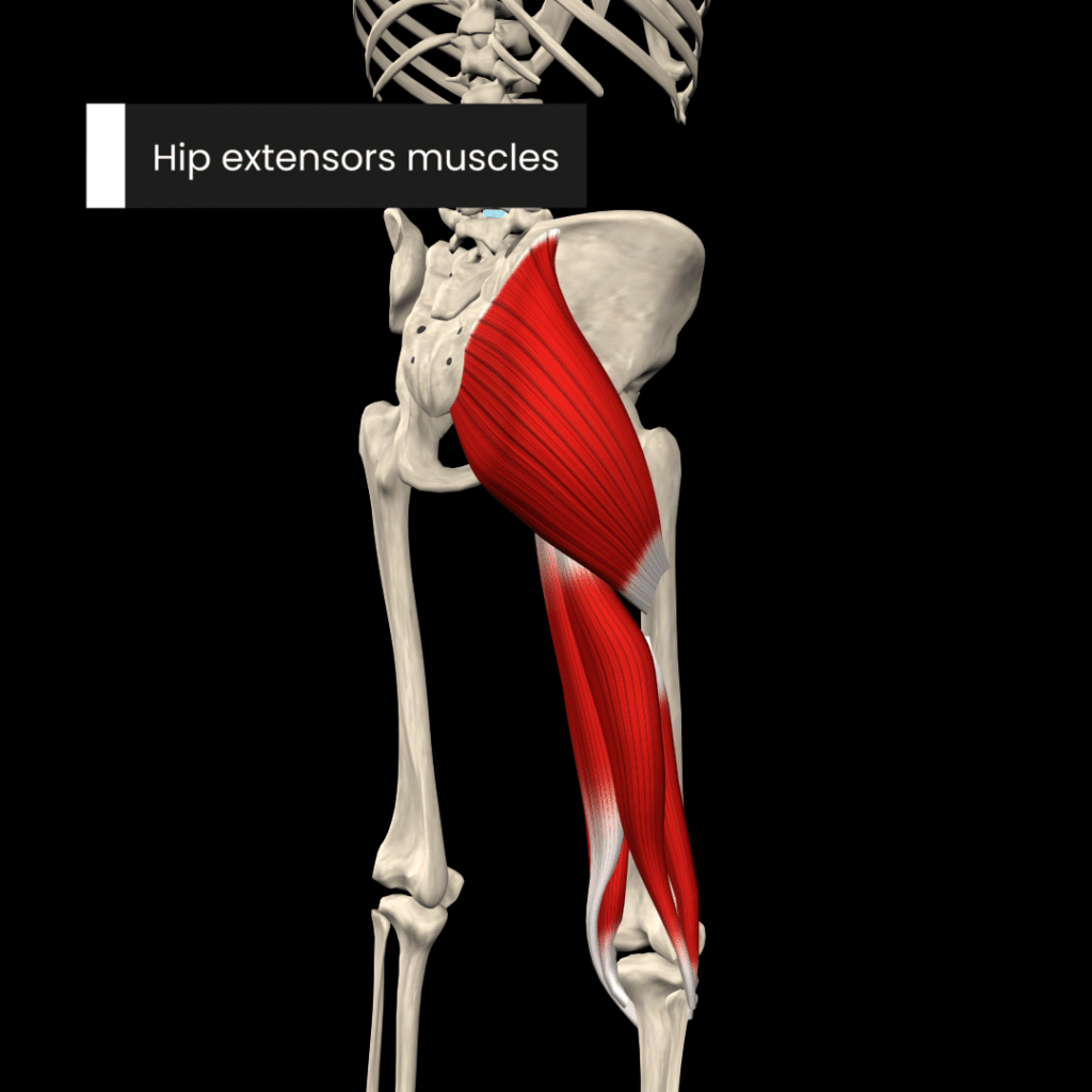
Hip extensors muscles
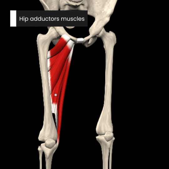
Hip adductors muscles
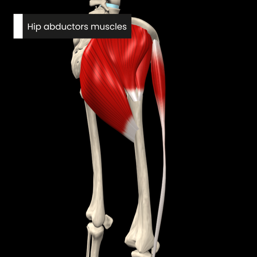
Hip abductors muscles
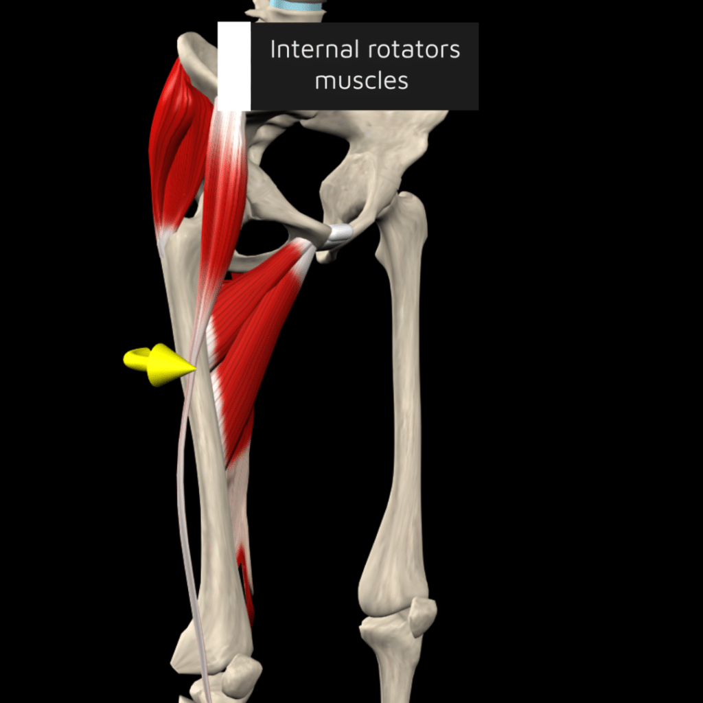
Hip internal rotator muscles
- Gluteus minimus
- Gluteus medius
- Tensor fasciae latae
- Adductor magnus (hamstring part)
- Adductor longus
- Adductor brevis
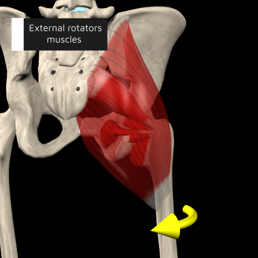
Hip external rotator muscles
- Gluteus maximus
- Gemellus superior
- Gemellus inferior
- Obturator externus
- Obturator internus
- Quadratus femoris
- Piriformis
In summary, the hip joint is an anatomical marvel that plays a crucial role in human movement and weight-bearing activities. Its ball-and-socket structure allows for a remarkable range of motion while the surrounding ligaments, muscles, and the acetabular labrum provide stability and protection. The arrangement of the joint capsule and ligaments enables secure weight distribution and mobility.
Check out our comprehensive Strength Training App (all anatomy content included in the strength app) to learn more about the fascinating world of hip anatomy and how each muscle contributes to movement. Our app provides detailed visualizations and interactive models to help you understand the complex interactions of hip muscles, ligaments, and bones.
Whether you’re a student eager to master anatomy, a professional looking to refresh your knowledge, or simply curious about how your body moves, our app offers a wealth of information at your fingertips.
Explore at your own pace, learn through engaging content, and discover the intricate mechanics of the hip joint that support your daily activities. Visit our website to access the app and start your journey into the dynamic world of human anatomy today!
Dive deeper into our content – Download our Strength Training App Today.
All anatomy content is included in the Strength Training App.
![]()
![]()
Reference:
- Glenister, R., & Sharma, S. (2023). Anatomy, bony pelvis and lower limb, hip. StatPearls Publishing.
- Netter. (2018). Atlas of Human Anatomy (7th ed.). Elsevier – Health Sciences Division.
- Lim, W. (2021). Clinical application and limitations of the capsular pattern. Physical Therapy Korea, 28(1), 13–17.
