Diastasis recti (DR) is a condition affecting many women, especially after childbirth. With various claims about the “best” exercises to fix this issue, it can be confusing to navigate what truly helps. This Muscle and Motion article provides a clear overview of diastasis recti.
We will explore the anatomy of the abdominal muscles and the role of hormonal changes during pregnancy and review different exercise strategies for effective recovery. We aim to offer practical, evidence-based advice to help manage and improve diastasis recti.
What is diastasis recti?
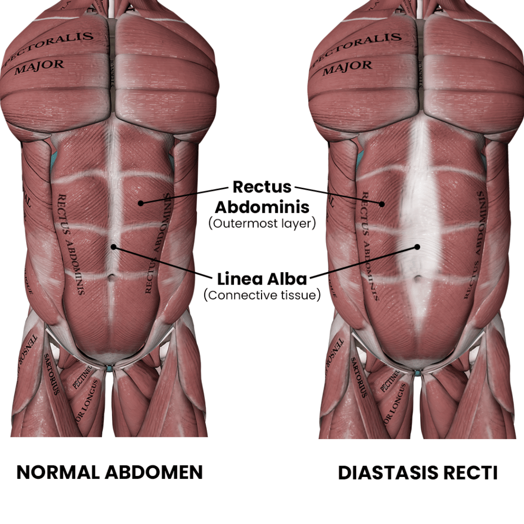
Diastasis recti occurs when the space between the left and right abdominal muscles widens. Typically, a separation of more than 2 cm is considered diastasis recti.
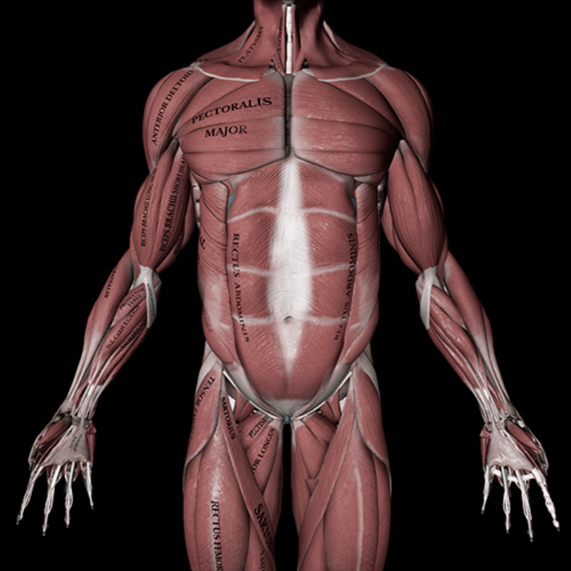
To understand why the space between the abdominal muscles increases, it’s essential first to understand their anatomy. The abdominal muscles consist of four main layers: the external oblique, internal oblique, transverse abdominis, and rectus abdominis, often known as the “six-pack” muscles. These muscles are connected by a central band of connective tissue called the linea alba, which helps keep your core stable.
During pregnancy, the body undergoes hormonal changes that allow the uterus to expand, which stretches the abdominal muscles, especially the rectus abdominis. Hormones such as relaxin, progesterone, and estrogen play a significant role in this process. These hormones help to loosen the connective tissue and prepare the body for childbirth. Additionally, the forward tilt of the pelvis and the curve in the lower back, which are also normal changes, can affect how these muscles support the organs. As the pregnancy progresses, the rectus abdominis muscles continue to stretch, which can naturally weaken them and lead to diastasis recti.[1]
Prevalence
Diastasis recti is a common condition that occurs during and after pregnancy but can also affect men and postmenopausal women, particularly those with age-related or abdominal weight gain.
Prevalence in women
The prevalence of diastasis recti in women changes over time:
33.1% at 21 weeks of pregnancy
60% at six weeks postpartum
45.4% at six months postpartum
32.6% at one year postpartum
These statistics indicate that while DR is very common shortly after childbirth, the prevalence decreases over time as some women’s bodies naturally heal. However, a significant percentage of women still experience DR up to a year after giving birth.
The impact of diastasis recti on functional problems
Diastasis recti is a condition that worries many women, but what does the research say about its effects on functional issues?
Two studies examined the relationship between DR and various functional problems. One study found no link between DR and lower back pain.[2] Another study examined whether DR was related to pelvic floor issues like stress urinary incontinence, fecal incontinence, and pelvic organ prolapse. The findings showed no connection between DR and these pelvic floor problems.[3]
However, some researchers suggest that changes in the abdominal muscles associated with DR may impair muscle strength, alter breathing patterns, and potentially lead to low back or pelvic girdle pain, as well as other pelvic floor disorders.[4-5]
Many women seek professional advice on exercises to help reduce the separation between their abdominal muscles. It’s not just about improving how their body functions — these women also want to enhance the structure and appearance of their abdominal area.
Enhancing recovery for postpartum diastasis recti
Natural recovery
Research shows that the natural recovery process of diastasis recti postpartum involves significant improvement in the gap between abdominal muscles over six months, particularly above and at the navel, with a reduction of about 2 cm.[6] Despite this progress, postpartum women often still exhibit a wider gap and weaker muscles compared to those who haven’t given birth. This difference highlights the need for targeted exercises, such as deep core muscle contractions, to enhance recovery and restore abdominal strength more effectively.
Exercises
Abdominal crunch exercise
Abdominal crunches, which involve lifting the head and chest while lying down, are often debated as a viable treatment for women with diastasis recti. Some fear they increase intra-abdominal pressure and worsen the gap. However, studies show that crunches can reduce the gap size for postpartum women. For instance, one study found a significant reduction in the gap during crunches compared to rest.[7]
Crunches vs. TA exercises
Another study comparing crunches to transversus abdominis (TA) exercises found crunches more effective in narrowing the gap at multiple points above and below the navel, particularly six weeks postpartum.[8]
Combined approach
A study examined the impact of crunches and transverse abdominis curls (crunches with an added contraction of the transverse abdominis) on diastasis recti in postpartum women.[9] Using ultrasound, researchers assessed the inter-recti distance (gap) in 26 women with diastasis recti and 17 control participants. The findings revealed that regular crunches were significantly more effective in reducing the gap compared to transverse abdominis curls, which had a lesser impact. However, the study concluded that while regular crunches are superior, incorporating transverse abdominis curls can further aid recovery. Thus, combining both exercises may enhance recovery outcomes for women with diastasis recti.
A 2023 systematic review of 16 trials involving 698 postnatal women found that abdominal exercises resulted in a modest reduction in the distance between the abdominal muscles compared to usual care, though this change was not clinically significant. Despite this, conservative interventions, including abdominal exercises, may still offer other physical and psychosocial benefits for managing abdominal separation.[10]
Progressive abdominal exercises
Since no single “best” abdominal exercise exists, this program is designed to gradually build core strength through a progressive approach. For postnatal exercise, it’s essential to include additional components such as aerobic activities and exercises targeting other muscle groups. This program focuses exclusively on abdominal exercises. For comprehensive strength training, please check out our blog posts: “Full-body Training Programs” and “First Steps in the Gym: A Workout Program for Beginners.” Additionally, you can find structured workout programs in our Strength Training App.
The program consists of different phases, ranging from basic to advanced exercises. You can choose the phase that matches your ability level.
Five-step progressive abdominal exercise program
Step 1: Establish basic core strength and stability.
Basic crunch: Engage your rectus abdominis by lifting your shoulders off the ground while lying on your back with your knees bent.
Pelvic tilt exercises:Control your lumbar spine by performing foundational pelvic tilts.
Wall plank with foam roller: Hold a plank position against a wall with a foam roller to stabilize your core.
Bird dog: Extend one arm and the opposite leg while maintaining a stable core to engage your transverse abdominis.
Step 2: Increase core strength and introduce more dynamic movements.
Sit up: Perform full range of motion sit-ups to engage the rectus abdominis.
Supine bent-leg raise: Lift your bent legs towards your chest while lying on your back to target the lower rectus abdominis.
Supine bicycles: Mimic a cycling motion with your legs while lying on your back, alternating elbow to knee to work the obliques.
Elbow plank: Hold a plank position on your elbows to build core stability and endurance.
Step 3: Enhance core strength and stability with more challenging exercises.
Bicycle crunch: Lie on your back with your knees bent. Perform a cycling motion with your legs while touching opposite elbow to knee to target your obliques.
Butterfly sit-up: Sit with the soles of your feet together and perform a sit-up to engage your core in a wider range of motion.
Reverse crunch: Lie on your back with your knees bent, lift your hips towards your chest, and work your lower abs.
Elbows plank on stability ball: Hold a plank position with your elbows on a stability ball to increase instability and challenge your core.
Step 4: Maximize core strength and introduce functional movements.
Hanging leg raise: Hang from a pull-up bar with arms straight. Lift your legs towards your chest, keeping them straight to work your rectus abdominis.
Weighted crunch: Lie on your back with your knees bent. Hold a weight plate and perform crunches to target your rectus abdominis.
Side plank: Hold a plank position on your side to engage your obliques and improve lateral stability.
Hollow body flutter kicks:Lie on your back in a hollow body position and perform flutter kicks to engage your lower abs and hip flexors.
Step 5: Achieve peak core strength and stability with advanced exercises.
Oblique crunch with straps: Use straps to perform an oblique crunch, adding resistance and instability to challenge your obliques.
Abs wheel rollout: Start on your knees with an ab wheel. Roll forward, extending your body while keeping your core engaged, then roll back.
Dragon flag: Lie on a bench, hold the edges, and lift your body off the bench except for your upper back, keeping a straight line from shoulders to feet.
Alternating toe touch: Lie on your back with your legs up. Reach hands to toes, lifting your shoulders to target your rectus abdominis and obliques.
Ever wondered what makes our anatomical animations so accurate and engaging? Click here to learn about our Quality Commitment and the experts behind our content.
At Muscle and Motion, we believe that knowledge is power, and understanding the ‘why’ behind any exercise is essential for your long-term success.
Let the Strength Training App help you achieve your goals! Sign up for free.
Reference:
Boissonnault, J. S., & Blaschak, M. J. (1988). Incidence of diastasis recti abdominis during the childbearing year. Physical Therapy, 68(7), 1082–1086.
Mota, P. G., Pascoal, A. G., Carita, A. I., & Bø, K. (2015). Prevalence and risk factors of diastasis recti abdominis from late pregnancy to 6 months postpartum, and relationship with lumbo-pelvic pain. Manual Therapy, 20, 200–205.
Bø, K., Hilde, G., Tennfjord, M. K., et al. (2017). Pelvic floor muscle function, pelvic floor dysfunction, and diastasis recti abdominis: Prospective cohort study. Neurourology and Urodynamics, 36, 716–721.
Skoura, A., Billis, E., Papanikolaou, D. T., Xergia, S., Tsarbou, C., Tsekoura, M., Kortianou, E., & Maroulis, I. (2024). Diastasis recti abdominis rehabilitation in the postpartum period: A scoping review of current clinical practice. International Urogynecology Journal, 35(3), 491–520.
Liaw, L. J., Hsu, M. J., Liao, C. F., et al. (2011). The relationships between inter-recti distance measured by ultrasound imaging and abdominal muscle function in postpartum women: A 6-month follow-up study. Journal of Orthopaedic & Sports Physical Therapy, 41(6), 435–443.
Liaw, L. J., Hsu, M. J., Liao, C. F., et al. (2011). The relationships between inter-recti distance measured by ultrasound imaging and abdominal muscle function in postpartum women: A 6-month follow-up study. Journal of Orthopaedic & Sports Physical Therapy, 41, 435–443.
Chiarello, C. M., McAuley, J. A., & Hartigan, E. H. (2016). Immediate effect of active abdominal contraction on inter-recti distance. Journal of Orthopaedic & Sports Physical Therapy, 46, 177–183.
Mota, P., Pascoal, A. G., Carita, A. I., & Bø, K. (2015). The immediate effects on inter-rectus distance of abdominal crunch and drawing-in exercises during pregnancy and the postpartum period. Journal of Orthopaedic & Sports Physical Therapy, 45, 781–788.
Chiarello, C. M., McAuley, J. A., & Hartigan, E. H. (2016). Immediate effect of active abdominal contraction on inter-recti distance. Journal of Orthopaedic & Sports Physical Therapy, 46, 177–183.
Benjamin, D. R., Frawley, H. C., Shields, N., Peiris, C. L., van de Water, A. T. M., Bruder, A. M., & Taylor, N. F. (2023). Conservative interventions may have little effect on reducing diastasis of the rectus abdominis in postnatal women – A systematic review and meta-analysis. Physiotherapy, 119, 54–71.