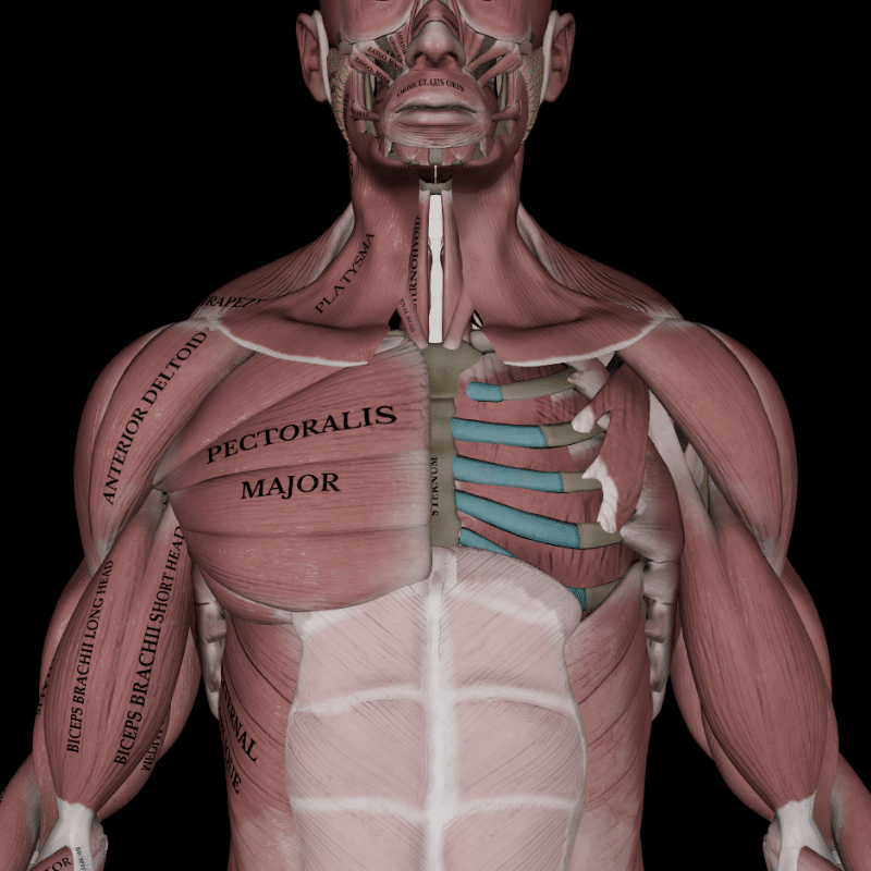
The pectoral muscles, a group of skeletal muscles that attach the upper limbs to the front and sides of the thoracic wall, are more than just anatomical structures. They are the unsung heroes of our daily activities, enabling us to perform tasks as simple as pushing a door or lifting a bag.
As some of the most prominent muscles of the upper torso, these muscles play a vital role in stabilizing the shoulder girdle and enabling a wide range of arm movements.
This detailed Muscle and Motion article will explore the anatomy of the pectoral muscles, their functions, and their importance in maintaining upper body strength and stability.
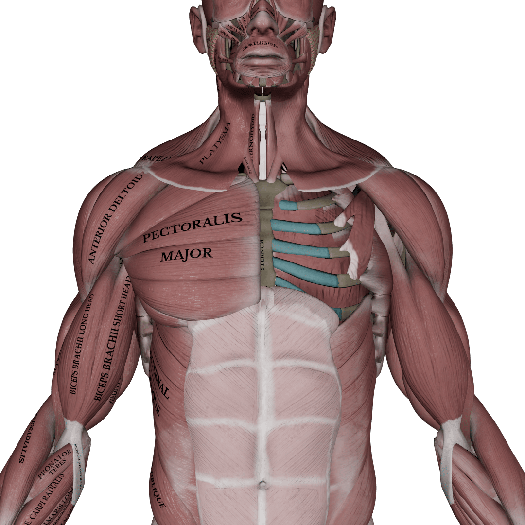
Anatomy of the pectoral muscles
The pectoral region contains several vital muscles, including the pectoralis major, pectoralis minor, subclavius, and serratus anterior. Each plays a unique role in shoulder and upper arm movement and stability.
Pectoralis major
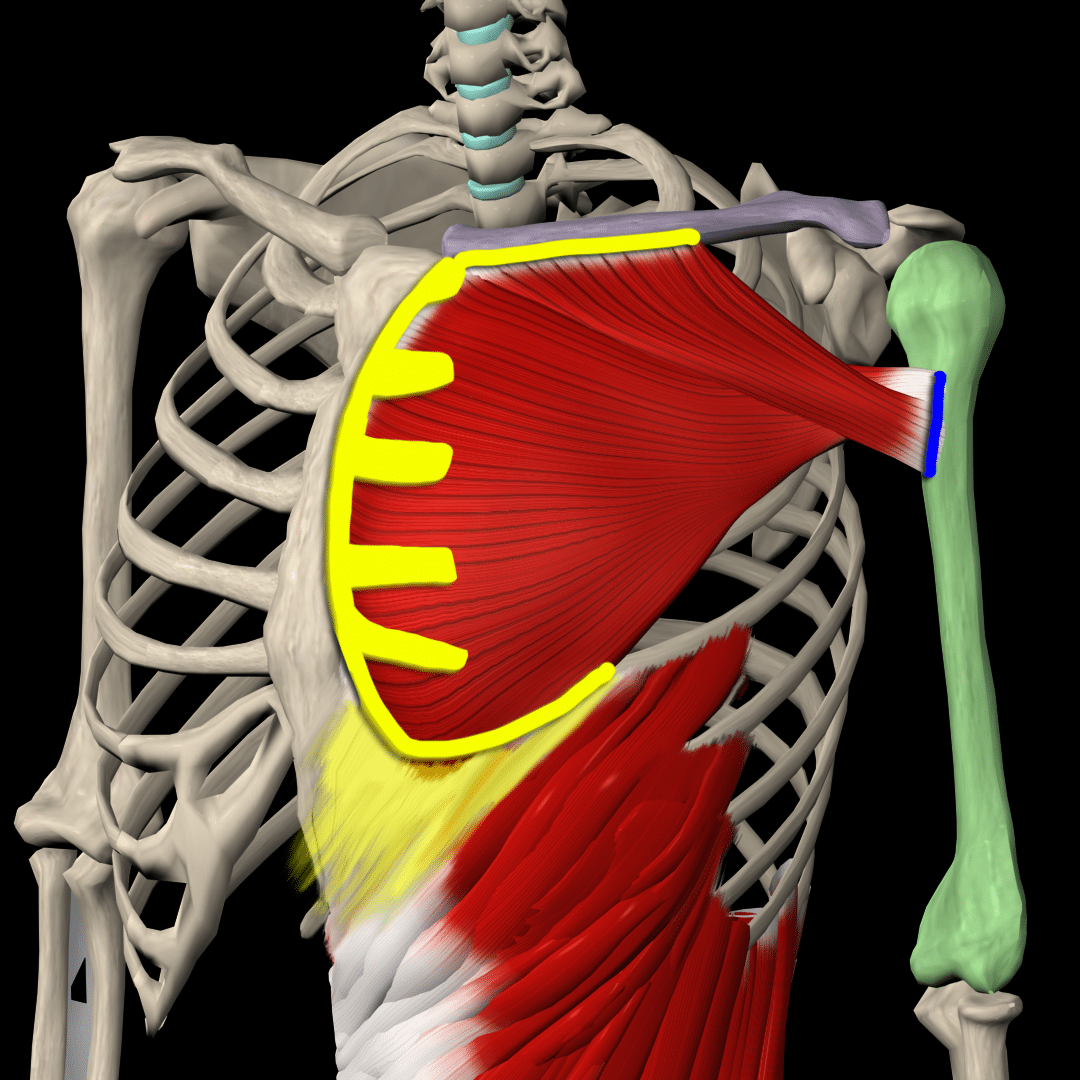
The pectoralis major is the anterior chest wall’s largest and most superior muscle, prominently positioned in the upper chest. This muscle consists of three distinct segments: the clavicular part, the sternocostal part, and the coastal part.
Origin (proximal attachment):
- Clavicular head: From the anterior surface of the medial half of the clavicle
- Sternocostal head: From the anterior surface of the sternum and the first seven costal cartilages
- Costal head: From the cartilage of the 6th rib and the aponeurosis of the external oblique muscle
Insertion (distal attachment):
- All three parts of the pectoralis major converge laterally, forming a broad tendon that inserts into the lateral lip of the intertubercular sulcus of the humerus.
Innervation:
- Lateral and medial pectoral nerves (C5-T1)
Functions:
The pectoralis major is responsible for several critical movements of the shoulder:
- Horizontal adduction
- Internal rotation
- ֿShoulder adduction
- Clavicular head: Flexes the shoulder when extended
- Sternocostal head: Extends the shoulder when flexed
- Assists in forced inhalation by pulling the rib cage forward to expand the chest
Pectoralis minor
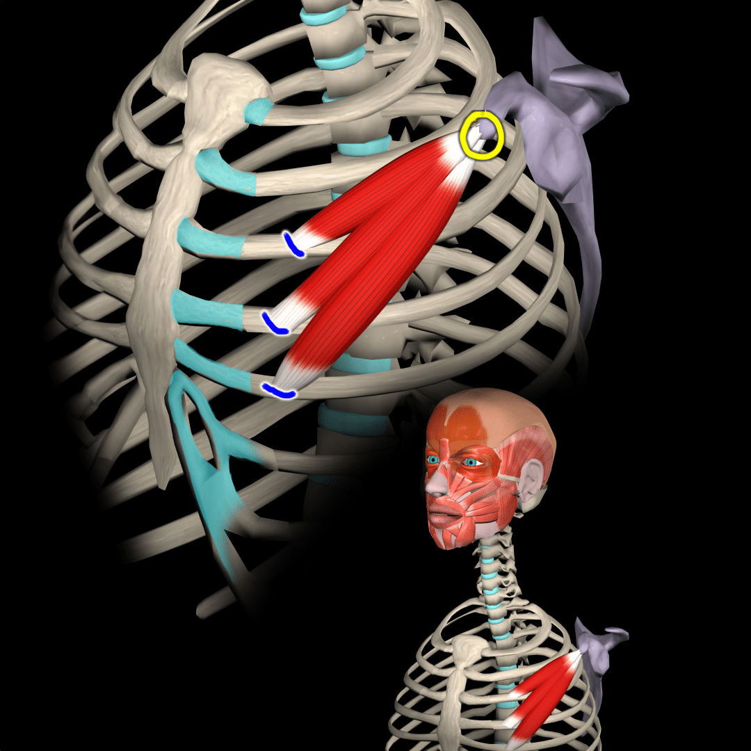
The pectoralis minor is a thin, triangular muscle beneath the pectoralis major, lying deep within the anterior chest wall. Though smaller, it plays a vital role in stabilizing and moving the scapula.
Origin (proximal attachment):
- From the anterior surfaces of the 3rd-5th, near their costal cartilages
Insertion (distal attachment):
- Attaches to the medial border and upper surface of the coracoid process of the scapula
Innervation:
- Medial pectoral nerve (C8-T1)
Functions:
The pectoralis minor is primarily involved in the following movements and functions:
- Scapular depression
- Protraction of the scapula
- Downward rotation of the scapula
- Assists in forced inhalation by elevating the ribs to expand the chest.
Subclavius
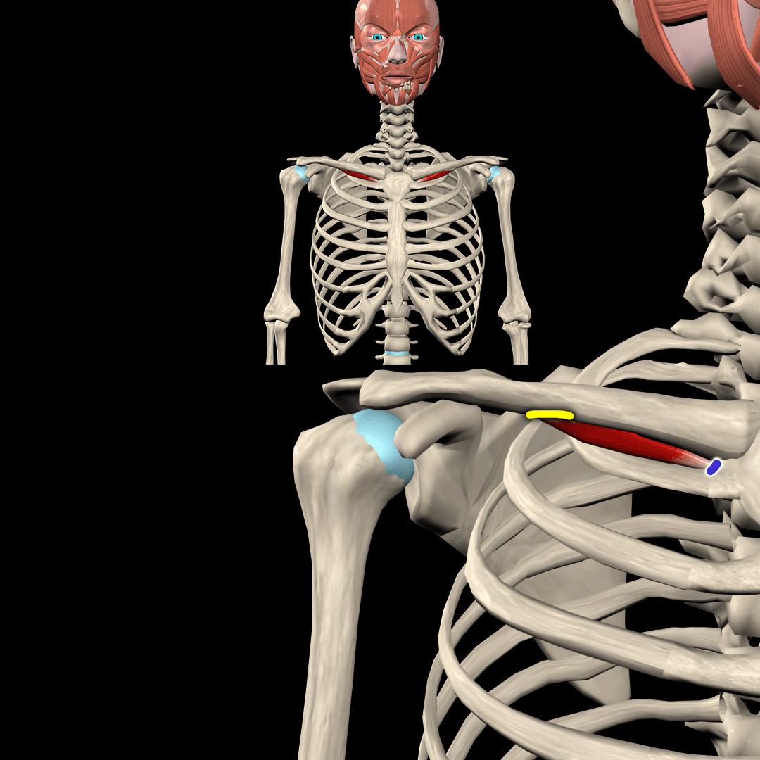
The subclavius is a small, cylindrical muscle located beneath the clavicle, lying below the pectoralis major. It serves a crucial role in stabilizing the clavicle and protecting underlying structures.
Origin (proximal attachment):
- From the first rib and its costal cartilage
Insertion (distal attachment):
- Attaches to the inferior surface of the middle third of the clavicle
Innervation:
- Nerve to subclavius (C5-C6)
Functions:
The subclavius muscle is involved in the following functions:
- Stabilizes the clavicle during shoulder movements
- Depresses the clavicle, moving it downward and forward
- Helps protect the brachial plexus and subclavian vessels under the clavicle during movements or trauma
Serratus anterior
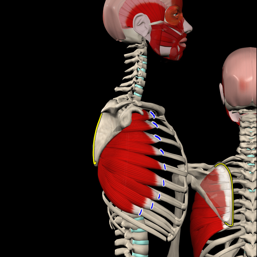
The serratus anterior is a fan-shaped muscle located on the lateral wall of the thorax, positioned between the rib cage and the scapula. It plays a key role in the movement and stabilization of the scapula.
Origin (proximal attachment):
- The outer surface of the upper 8 or 9 ribs
Insertion (distal attachment):
- Attaches to the costal surface of the medial border of the scapula
Innervation:
- Long thoracic nerve (C5, C6, C7)
Functions:
- Scapular protraction
- Scapula upward rotation
- Prevents winging of the scapula and ensures stable shoulder movement
- Assists in ventilation by lifting the rib cage during inhalation when the scapula is fixed, aiding in breathing
Strengthening the muscles of the pectoral region
To strengthen the muscles in the pectoral region, you must include a variety of exercises, as these muscles are responsible for multiple movements. Here are the key exercise types to add to your workout routine:
Compound pressing movements
Compound pressing exercises, such as bench presses and push-ups, are fundamental for building overall chest strength. These movements primarily target the pectoralis major and also engage supporting muscles like the deltoids and triceps. By adjusting the angle of the press (flat, incline, or decline), you can emphasize different areas of the pectoralis major, focusing on the upper, middle, or lower fibers.
Isolation movements
Isolation exercises, like cable flies and pec deck flies, focus specifically on the pectoralis major by directly targeting the muscle fibers and reducing the involvement of other muscle groups.
Scapular stability exercises
While pushing exercises like bench presses and shoulder presses also engage muscles like the serratus anterior, pectoralis minor, and subclavius, scapular stability exercises emphasize these muscles more. For example, scapular push-ups and scapular protraction specifically target the serratus anterior. These exercises are excellent for strengthening weak links, enhancing scapular stability, and helping to manage shoulder pain during pushing movements.
In this blog, we explored the anatomy and functions of the pectoral muscles, including the pectoralis major, pectoralis minor, subclavius, and serratus anterior, highlighting their crucial role in providing strength and stability to the shoulder girdle and enabling a wide range of arm movements.
We also offered a variety of exercises to specifically target and strengthen these muscles, enhancing overall upper-body performance.
Understanding these muscles’ functions is essential for students, trainers, coaches, and anyone involved in sports. Our anatomy section, featured in all our apps, including the Strength Training App, offers a dynamic and interactive learning experience with detailed 3D models, instructional videos, and comprehensive explanations to help you easily understand each muscle’s origin, insertion, innervation, and actions, ultimately deepening your knowledge of human anatomy.
Have you ever wondered what makes our anatomical animations so accurate and engaging? Click here to learn about our Quality Commitment and the experts behind our content.
At Muscle and Motion, we believe that knowledge is power, and understanding the ‘why’ behind any exercise is essential for your long-term success.
Let the Strength Training App help you achieve your goals! Sign up for free.
