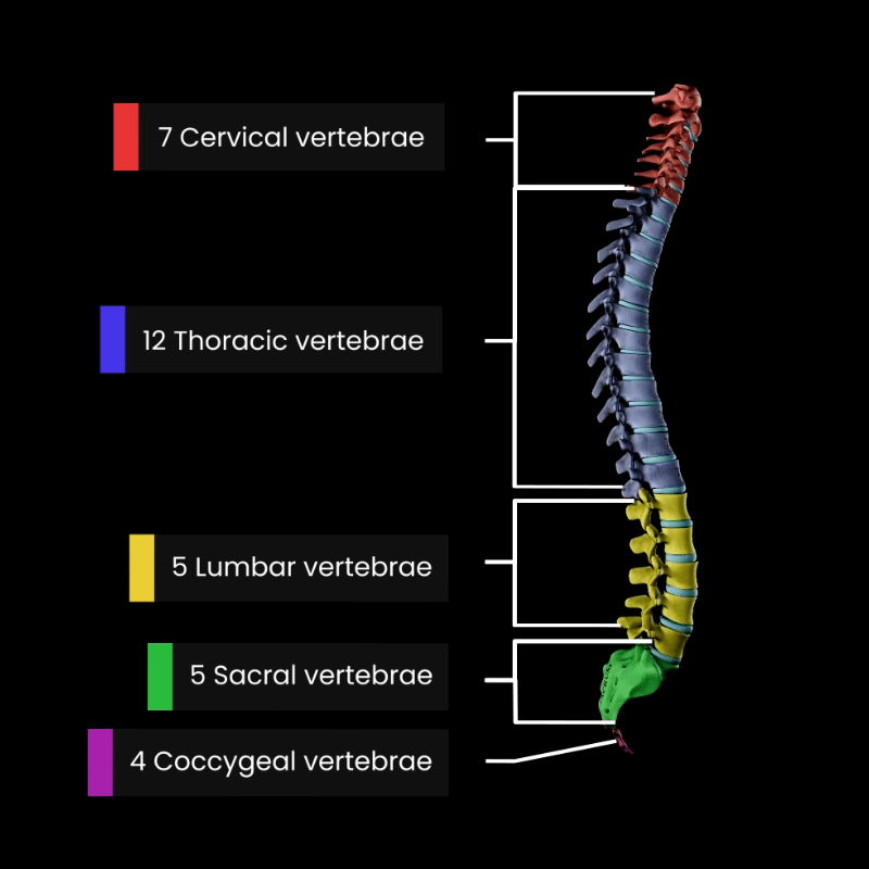
The vertebral column, commonly known as the spine, is a vital structure that supports and protects the human body. It consists of 33 individual bones, called vertebrae, separated by intervertebral discs, which provide a perfect balance of flexibility and stability.
The spine is divided into five distinct regions, each with its own specialized function:
- 7 cervical vertebrae (neck region)
- 12 thoracic vertebrae (mid-back, connected to the rib cage)
- 5 lumbar vertebrae (lower back)
- 5 sacral vertebrae (fused to form the sacrum)
- 4 coccygeal vertebrae (fused to form the coccyx or tailbone)
In this Muscle and Motion article, we’ll explore the intricate structure of the vertebral column, exploring the unique features of each region and the characteristics that make this structure essential for movement, support, and protection.
The general structure of a vertebra
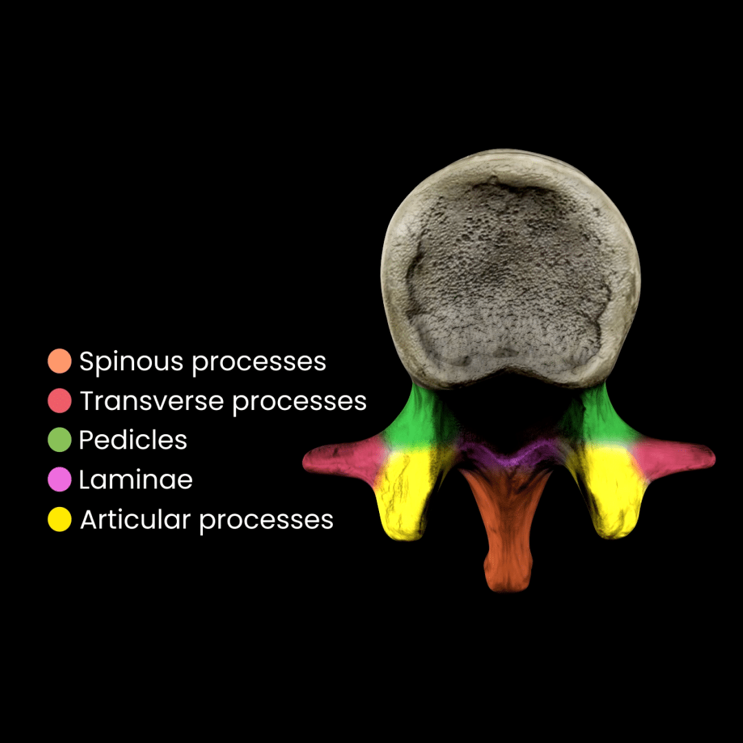
Each vertebra comprises two primary parts: the anterior vertebral body and the posterior vertebral arch, which play vital roles in the vertebra’s function and structure.
The vertebral body
The vertebral body is the primary weight-bearing structure. It is situated at the front of the vertebra and gradually increases in size in the lower spine to accommodate the greater load it supports. The vertebral body’s top (superior) and bottom (inferior) surfaces are coated with hyaline cartilage. Intervertebral discs separate the vertebral body from adjacent vertebrae, aiding in shock absorption and flexibility.
The vertebral arch
The vertebral arch forms the vertebra’s back (posterior) and sides (lateral). Together with the vertebral body, it encloses the vertebral foramen. This central opening aligns with those of adjacent vertebrae to create the vertebral canal, which houses and protects the spinal cord.

Several bony projections characterize the vertebral arch:
- Spinous and transverse processes: Serve as attachment points for muscles and ligaments, aiding in movement and stability.
- Pedicles and laminae: Form the base and roof of the vertebral arch, respectively.
- Articular processes: Connect adjacent vertebrae and contribute to spinal flexibility.
This intricate structure allows the vertebrae to provide robust support and flexibility for movement and stability.
Breaking down the spinal regions
The vertebral column, a cornerstone of human anatomy, comprises five distinct regions, each tailored to specific functions and characterized by unique structural features. These regions work together to provide stability and flexibility, support the body, and protect the spinal cord.
Cervical vertebrae
The cervical region is located in the neck and is designed for mobility and support of the head. Its seven vertebrae exhibit the following characteristics:
- Small vertebral bodies accommodate the reduced weight they bear.
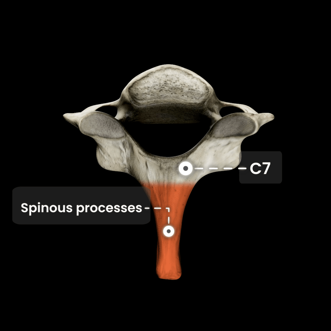
- Bifid spinous processes, where the spinous process splits into two parts, except in C1 and C7.
- Transverse foramina are small openings that allow for the passage of vertebral arteries.
A triangular vertebral foramen is distinct from this region.
Certain vertebrae have unique characteristics that are important to recognize. These include:
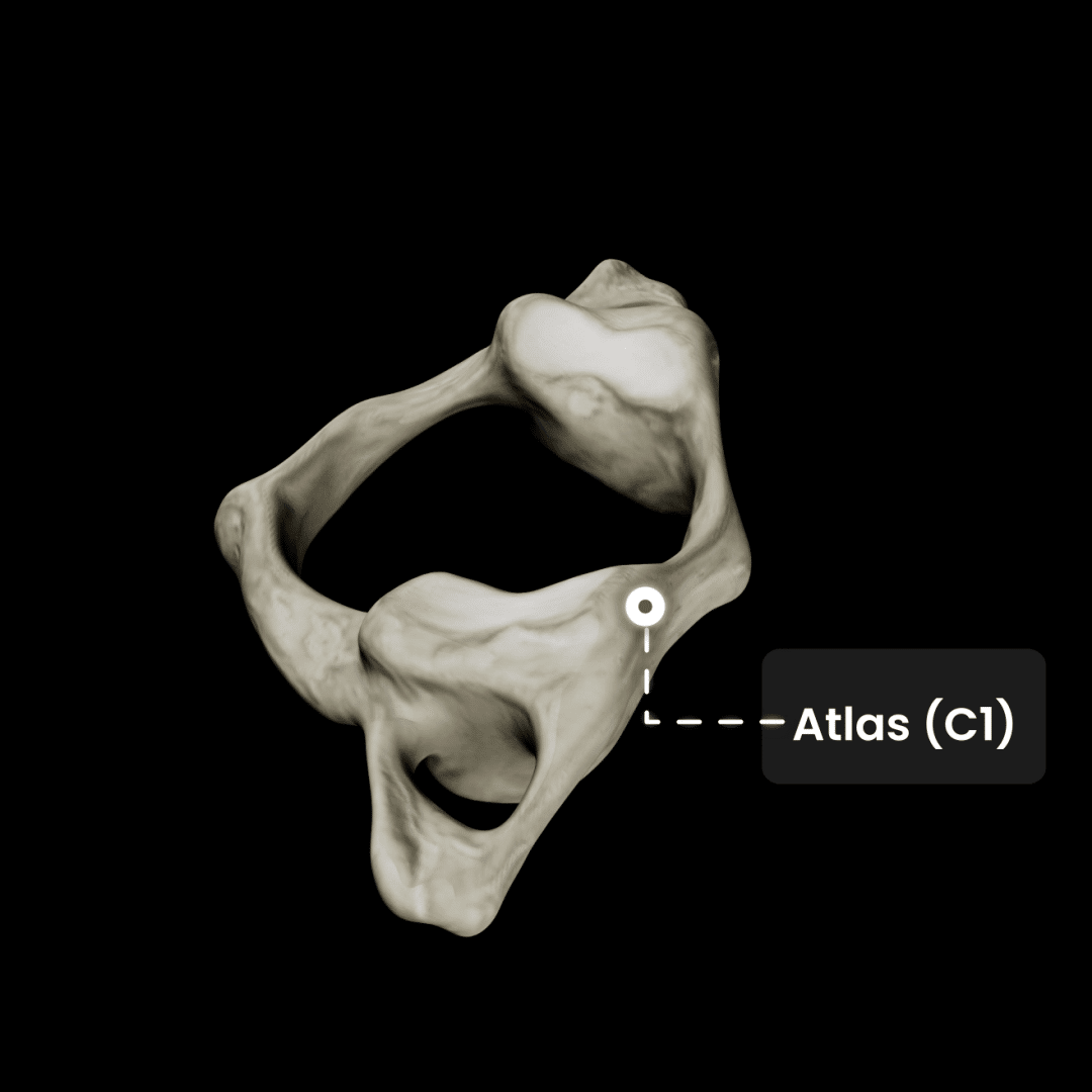
- Atlas (C1): The first vertebra supports the skull and allows nodding movements. It is ring-shaped and uniquely lacks a vertebral body or spinous process.
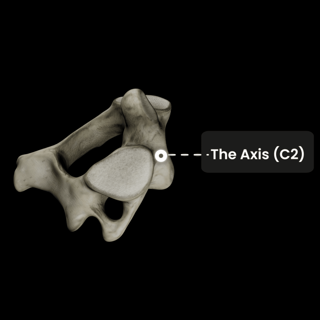
- Axis (C2): Known for its odontoid process (dens), this vertebra acts as a pivot, enabling side-to-side head rotation.
- C7 (vertebra prominens): Recognized for its prominent spinous process, it also resembles a thoracic vertebra, acting as a transitional vertebra between the cervical and thoracic regions.
Thoracic vertebrae
The thoracic spine consists of 12 vertebrae, each uniquely adapted for rib articulation and the protection of vital organs. These vertebrae feature:
- Demi facets, allowing ribs to attach at two points on each vertebra.
- Costal facets on the transverse processes for rib articulation.
- Obliquely angled spinous processes that overlap like shingles to increase stability.
- A circular vertebral foramen, differing from the triangular shape in the cervical and lumbar regions.
This region anchors the rib cage, providing a flexible framework for respiration and upper body movement.
Lumbar vertebrae
The lumbar spine comprises five vertebrae to support most of the body’s weight. These vertebrae are the largest and strongest in the spine. Key features include:
- Large vertebral bodies are designed for weight-bearing.
- Transverse foramina, costal facets, and bifid spinous processes are absent in other regions.
- A triangular vertebral foramen, similar to the cervical region, but with a design optimized for lumbar function.
This region is vital for the mobility and stability of the lower back and torso.
Sacrum and coccyx
- Sacrum: A triangular bone composed of five fused vertebrae, it connects the spine to the pelvis at the sacroiliac joint.
- Coccyx (tailbone): Formed by four fused vertebrae, the coccyx is a remnant of evolutionary history and provides attachment for ligaments and muscles.
Joints and ligaments
The vertebrae articulate at joints supported by robust ligaments that maintain stability and enable movement:
- Anterior and posterior longitudinal ligaments reinforce the vertebral bodies.
- Ligamentum flavum connects adjacent vertebrae and helps maintain posture.
- Interspinous and supraspinous ligaments stabilize the spinous processes.
- Intertransverse ligaments link transverse processes, contributing to lateral stability.
In summary, the vertebral column is a remarkable anatomical structure, divided into five regions: cervical, thoracic, lumbar, sacral, and coccygeal. Each region is uniquely designed to fulfill its specific role, from the intricate movement capabilities of C1 and C2 in the neck to the robust weight-bearing function of the lumbar vertebrae in the lower back.
A clear understanding of these regions highlights the spine’s dual role in supporting and enabling movement while protecting the spinal cord. For a deeper, interactive exploration of spinal anatomy, check out the Muscle and Motion Strength Training App and see how these structures work seamlessly together.
Have you ever wondered what makes our anatomical animations so accurate and engaging? Click here to learn about our Quality Commitment and the experts behind our content.
At Muscle and Motion, we believe that knowledge is power, and understanding the ‘why’ behind any exercise is essential for your long-term success.
Let the Strength Training App help you achieve your goals! Sign up for free.
All anatomy content is included in the Strength Training app.
![]()
![]()